Nuclear medicine is a specialized branch of medical imaging that uses small amounts of radioactive materials (radiopharmaceuticals) to diagnose and treat various diseases.
It plays a crucial role in modern healthcare by enabling early detection, accurate diagnosis, and targeted treatments for conditions such as cancer, cardiovascular diseases, and neurological disorders.
Techniques like Positron Emission Tomography (PET), Single Photon Emission Computed Tomography (SPECT), and radionuclide therapy have revolutionized patient care by providing detailed functional imaging beyond what conventional radiology can offer.
A well-designed nuclear medicine unit is essential for ensuring patient and staff safety, operational efficiency, and compliance with strict radiation protection regulations.
Proper planning minimizes radiation exposure, optimizes workflow, and enhances patient experience while meeting international guidelines set by regulatory bodies such as the International Atomic Energy Agency (IAEA) and the Nuclear Regulatory Commission (NRC).
If you want to know about the Types of slabs or Permeable concrete or Islamic architecture, please click the link.
1) Introduction
The Nuclear Medicine Unit provides facilities for the administration of radiopharmaceutical agents to patients and patient imaging for diagnostic purposes and for treatment. The Nuclear Medicine Unit may be provided within the Medical Imaging Unit or as a freestanding Unit.
The Unit may or may not include a Radiopharmacy Laboratory. The size of a unit in terms of numbers and type of cameras will be determined by the service plan and clinical needs.
2) Planning of Nuclear Medicine Unit
i) Model of Care
The model of care will depend on level of services provided as defined in the service plan and the presence or otherwise of PET as a sub-component of the Nuclear Medicine Unit.
In large centres, it will be a discrete unit. If there are only one or two gamma cameras, it may be a discrete sub-unit of Medical Imaging.
All units will have a Hot Laboratory (Hot Lab). Large centres may or may not include a Radiopharmacy Laboratory that will prepare its own radiopharmaceuticals for general use.
ii) Planning Models
Location
A ground floor site is preferred but if this cannot be achieved, consideration should be given to units above, below and adjoining the proposed location with regard to radiation shielding requirements, the weight of equipment and associated shielding and access for equipment and radioactive isotopes. The Unit should not act as a thoroughfare to other units of the healthcare facility.
Unit layout
Staff and patient flows in the Unit are critical to ensure that patients, staff and visitors are not exposed to radiation as a result of travel through or adjacent to areas occupied by dosed patients and scanning rooms. Effective layout can also reduce the need for costly radiation shielding.
Layout should address the need for separation of areas particularly patient and staff corridors and entry areas for outpatients and inpatient on beds / trolleys.
If provided, the Bone Density Room should be located near the entry to the Nuclear Medicine Unit to ensure patients do not unnecessarily cross areas of radioactivity. The Bone Densitometry room should be located away from dosed patients by distance or shielding to avoid interference to the Bone Density Unit from high ambient radiation levels.
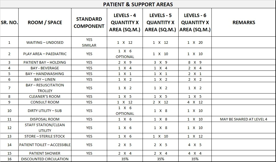
iii) Functional Areas
The Nuclear Medicine Unit consists of the following functional areas:
- Reception / Administration
- Waiting areas for outpatients and inpatients, including toilets
- patient holding, observation and recovery area
- treatment areas including gamma camera rooms, specialised scanning imaging rooms (SPECT, PET, PET/CT, bone densitometry), stress testing facilities
- support areas including utilities, staff station
- Hot Lab / Radioactive Waste Store
- staff areas including offices and amenities
- teaching and research facilities (Tertiary Centres)
Patient waiting
Waiting areas should allow separation of dosed and undosed patients, particularly as some patients may need to wait for 45 minutes after dosing for uptake. It is also preferable to separate dosed patients from relatives and visitors to the unit which may include young adults, pregnant women and children.
Dosed patients should have access to drinking water and toilet facilities without having to access general waiting areas. Outpatients should be separated from inpatients for privacy reasons with separate entrances.
Patient holding, Observation and Recovery area
An area will be required for patient holding, recovery and observation including the following:
- a dedicated inpatient entry
- curtained bed / trolley bays for holding, observation and recovery; the configuration of the overall space should permit both dosed and un-dosed patients to be held safely
- a small staff station with hand basin
- support rooms including Dirty Utility, Linen Bay, Sterile Stock Store
- Resuscitation trolley (trolley may be located near the Stress Testing Room).
Gamma camera
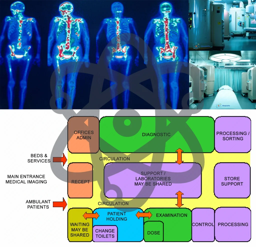
The gamma camera is a device used in Nuclear Medicine to image gamma radiation emitting radioisotopes to view and analyse images of the human body or the distribution of medically injected, inhaled, or ingested radionuclides emitting gamma rays, producing a two dimensional image.
The gamma camera consists of one or more flat crystal planes (detectors) optically coupled to an array of photomultiplier tubes, the assembly is known as a “head”, mounted on a gantry.
The gantry is connected to a computer system that both controls the operation of the camera as well as acquisition and storage of acquired images. The gamma camera room will require a control area and radiation screening as assessed by AERB Consultants.
Single photon emission computed tomography (SPECT)
SPECT is a nuclear medicine tomographic imaging technique using gamma rays, similar to the conventional gamma camera planar imaging system that is able to provide true 3D information. This information is typically presented as cross-sectional slices through the patient, but can be freely reformatted or manipulated as required.
To acquire SPECT images, the gamma camera is rotated around the patient. Projections are acquired at defined points during the rotation, typically every 3-6 degrees.
In most cases, a full 360 degree rotation is used to obtain an optimal reconstruction. The time taken to obtain each projection is also variable, but 15-20 seconds is typical. This gives a total scan time of 15-20 minutes.
A SPECT camera may be combined with a computerised tomography (CT) unit to form a hybrid system and fusion imaging of the physiology and anatomy of the area/s being scanned. SPECT/CT requires a separate control room and radiation screening in accordance with CT requirements.
Viewing and reporting area
A dedicated room with dimmable lighting will be required for viewing and reporting on scans. Each workstation should accommodate imaging screens, computers for access to imaging and patient information systems, writing and shelving space for reference materials.
The number of reporting stations will depend on service level, number of scanning rooms and the staff establishment.
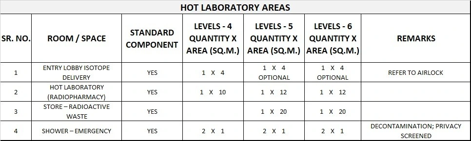
Hot Lab / Dispensary and Radioactive waste store
Radioactive radiopharmaceuticals are stored and prepared ready for administration to the patient in the Hot Lab. A lead screen barrier is required for the dispensary area.
The Dispensary should be located adjacent to the patient dosing room. A radioactive waste storage area may also be incorporated into or adjacent to this space.
Provide radiation shielding as advised by AERB Consultants. The Waste Store will require a sink and basin with hands-free taps for hand washing and equipment decontamination.
Radiopharmacy
The Radiopharmacy is used for preparation, compounding, quality control and dispensing of radiopharmaceuticals for diagnosis and treatment. Radiopharmaceuticals are radioactive isotopes attached to pharmaceutical substances.
Only designated units will have an in-house Radiopharmacy laboratory where cold kits are prepared; these may be used in-house or supplied to other Nuclear Medicine Units.
Many nuclear medicine units (e.g. private practices) may receive a daily delivery of the radiopharmaceutical already prepared and dispensed as individual patient doses.
Other isotopes / radionuclides (e.g. gallium, thallium) are delivered weekly or monthly as required, pre-packaged into individual doses for dispensing.
Storage – Equipment & Supplies
Storage is required for:
- collimators and scanning phantoms, within the scanning rooms.
- mobile equipment such as wheelchairs, trolleys, lifters and ultrasound scanners, may be located in equipment bays
- Technegas unit and large argon cylinder/s that may be located in an equipment bay ; the Technegas unit and trolley is taken to patients in holding bays or in the camera rooms for patient to inhale Tc99m
- Medical consumables and smaller equipment items
- sterile stock
- stationery
- records/ files.
iv) Functional Relationships
The Nuclear Medicine Unit should be located with ready access to the Medical Imaging Unit, PET Unit if provided, Emergency Unit, Operating Unit and Critical Care areas. It requires easy access for ambulant patients and beds/ stretchers.
3) Design of Nuclear Medicine Unit
i) Construction Standards
Construction Standards for a Nuclear Medicine Unit include the following:
- Flooring shall be adequate to meet load requirements for equipment, patients, and personnel.
- Floors and walls should be constructed of materials that are easily decontaminated in case of radioactive spills.
- Walls should contain necessary support systems for either built-in or mobile oxygen and vacuum, and vents for radioactive gases.
- Provision for cable trays, ducts or conduits should be made in floors, walls, and ceilings as required.
- Ceiling height should be a minimum of 3 metres.
- Ceiling mounted equipment should have properly designed rigid support structures located above the finished ceiling.
- A lay-in type ceiling should be considered for ease of installation, service and future remodelling.
ii) Environmental Considerations
Acoustics
Acoustic treatment will be required to the following areas:
- SPECT/CT scanning rooms (hybrid units may be noisy)
- Viewing / reporting room
- Consulting rooms
Refer also to acoustic requirements in “Acoustic Solutions for Healthcare Facilities”
Natural light
Natural light is desirable in all patient areas, staff room and staff offices. Lighting level in reporting rooms needs to be adjustable.
External windows provided in scanning and uptake rooms should be assessed by an AERB Consultant for shielding requirements.
iii) Safety and Security
The Nuclear Medicine Unit shall include a safety shower with an eyewash station for use in the event of radioactive spills.
iv) Finishes
Floor finishes and junctions should be smooth, impervious and non-absorbent in case of radiation spills.
v) Building Service Requirements
Radiation Protection
Plans and specifications will require assessment for radiation protection by an AERB consultant, as required by the appropriate state authorities. The radiation protection assessment will specify the type, location and amount of radiation protection required according to the final equipment selections and layout.
Radiation protection requirements shall be incorporated into the final specifications and the building plans. Radiation shielding will be required to a number of areas as advised by AERB Consultants including:
- Reception and rooms adjacent to dosed patient rooms
- Dosing/ Consult Exam rooms
- Hot Lab/ Dispensing room/ Radiopharmacy
- Pre-scan uptake rooms/ dosed waiting areas, patient toilets
- Cardiac Stress Testing Room
- Scanning Room/s
- Post scanning waiting areas
- Bone Densitometry Room
Hydraulic services
Ceiling spaces above gamma cameras and specialty scanning units should not be used for hydraulic services or air-conditioning ducts, to avoid damage to equipment from leakages.
The need for delayed holding tanks within the Nuclear Medicine Unit will require assessment by the AERB Consultant.
Mechanical services
Additional cooling and ventilation will be required to Scanning Rooms and associated computer equipment rooms as the equipment is sensitive to excessive ambient heat.
Some scanners may require chilled water for cooling. Large temperature changes (greater than 400C per hour) within scanning rooms need to be avoided to reduce the risk of crystal fracture in gamma cameras.
Additional air extraction or exhaust may be required to Camera Room/s where ventilation agents such as Technegas are administered.
General air conditioning inpatient and staff areas needs to be adjustable for patient and staff comfort; the temperature of the Unit should not exceed 25°C. Smoke detectors in treatment rooms should be sensitive to radiation. Hot Lab room air should be negatively pressured and exhausted, not recirculated.
The Hot Lab may include a fume cabinet which will require exhausting. Rooms in which Technegas is used should be negatively pressured to the rest of the Unit.
4) Components of the Unit
The Nuclear Medicine Unit will contain a combination of Standard Components and Non- Standard Components, according to the Level of Service.
Provide Standard Components to comply with details in the Standard Components described in these Guidelines. Refer also to Standard Components Room Data Sheets.
i) Non-Standard Components
SPECT and SPECT/CT Scanning room
Description and Function – The SPECT and SPECT/CT Scanning rooms will be used for patient imaging procedures using a SPECT camera or combined SPECT/CT hybrid system. Installation of equipment should be in accordance with manufacturer’s recommendations. Room size may vary according to the equipment selected
Location and Relationships – Scanning rooms require ready access from dosing rooms and dosed patient waiting areas. Scanning rooms may be collocated with shared Control rooms to enable monitoring of two rooms simultaneously.
Considerations
- Floor structure should support the equipment weight.
- Uninterruptible power supply is required to the cameras and associated computer modules to prevent data loss and/or damage during power surges or loss of supply
- Power to patient areas shall be body protected to protect against electric shock
- Individual room temperature and humidity control is required.
- Lighting should be placed to avoid lights shining directly into the patient’s eyes and should be dimmable.
- Radiation shielding will be required according to assessment by the AERB Consultant
- Bed / trolley access is required to the room
- Fixtures, fittings and equipment within the room will include:
- Collimator rack/s for a range of collimator sizes should be included; the collimator is a directional guide; the size and length of the collimator holes determine which gamma rays reach the detector in the camera; collimator racks vary according to the model / level of Gamma Camera.
- Patient ECG monitoring may be required in the room
- CCTV camera may be included (optional in SPECT/CT room).
- Lead apron rack and aprons will be required in the room or immediately adjacent.
- hand basin – Type B with paper towel and soap fittings
- Television, ceiling or wall mounted is optional.
- Bench and shelving for preparation and storage may be provided.
Services will include:
- medical gases – oxygen, suction, medical air on service panel
- nurse and emergency call system
- power outlets for patient equipment on the medical services panel and additional power on all walls
- computer data points on patient services panel, near gamma camera unit and in control areas.
Store – Radioactive waste
Description and Function – This room will store radioactive waste materials.
Location and Relationships – Ready access to the Hot Laboratory is required.
Considerations – Lead lining is required to ensure safe protection of radioactive materials.
Bone densitometry room
Description and Function – Bone densitometry is a non-invasive procedure using a special x-ray scanning machine to determine bone density or strength. It is used to identify those at risk of developing osteoporosis and to monitor change in bone density.
Location and Relationships – The bone densitometry room maybe located within the radiology or nuclear medicine
department.
Considerations – The room may have radiation shielding to walls and/or glazing as advised by AERB Consultant.
5) Schedule of Accommodation
Nuclear Medicine Unit Generic Schedule of Accommodation
Schedule of Accommodation for a Nuclear Medicine Unit for Level 4-6
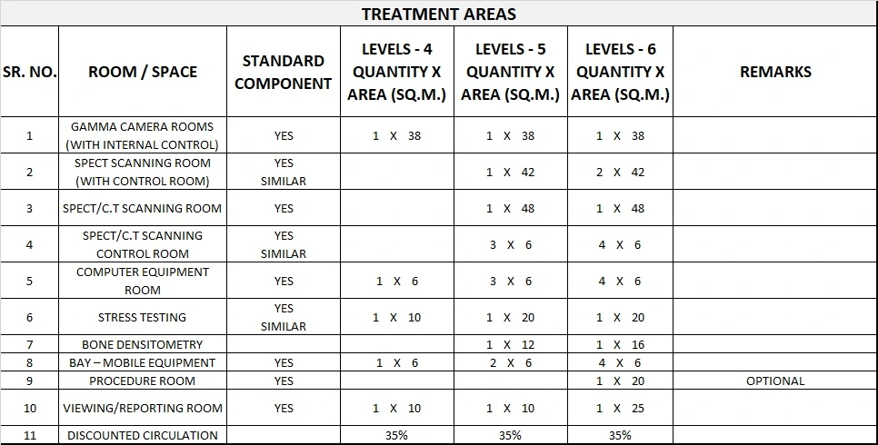
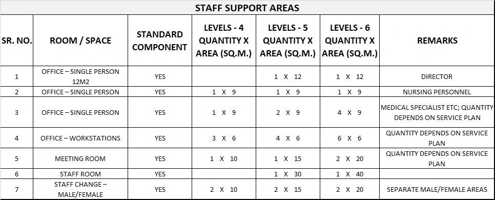
Please note the following:
- Areas noted in Schedules of Accommodation take precedence over all other areas noted in the FPU.
- Rooms indicated in the schedule reflect the typical arrangement according to the Role Delineation.
- Exact requirements for room quantities and sizes will reflect Key Planning Units identified in the service plan and the policies of the Unit.
- Room sizes indicated should be viewed as a minimum requirement; variations are acceptable to reflect the needs of individual Unit.
- Office areas are to be provided according to the Unit role delineation and staffing establishment.
- Staff and support rooms may be shared between Functional Planning Units dependant on location and accessibility to each unit and may provide scope to reduce duplication of facilities.
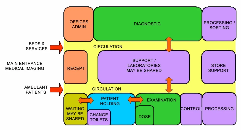
6) Functional Relationship Diagram
Nuclear Medicine Unit Functional Relationship Diagram