Medical imaging plays a crucial role in modern healthcare by enabling accurate diagnosis, treatment planning, and disease monitoring. It involves various imaging technologies such as X-ray, MRI, CT scans, ultrasound, and PET scans, each serving distinct medical purposes.
These imaging techniques help healthcare professionals detect abnormalities, guide surgeries, and track the progress of treatments.
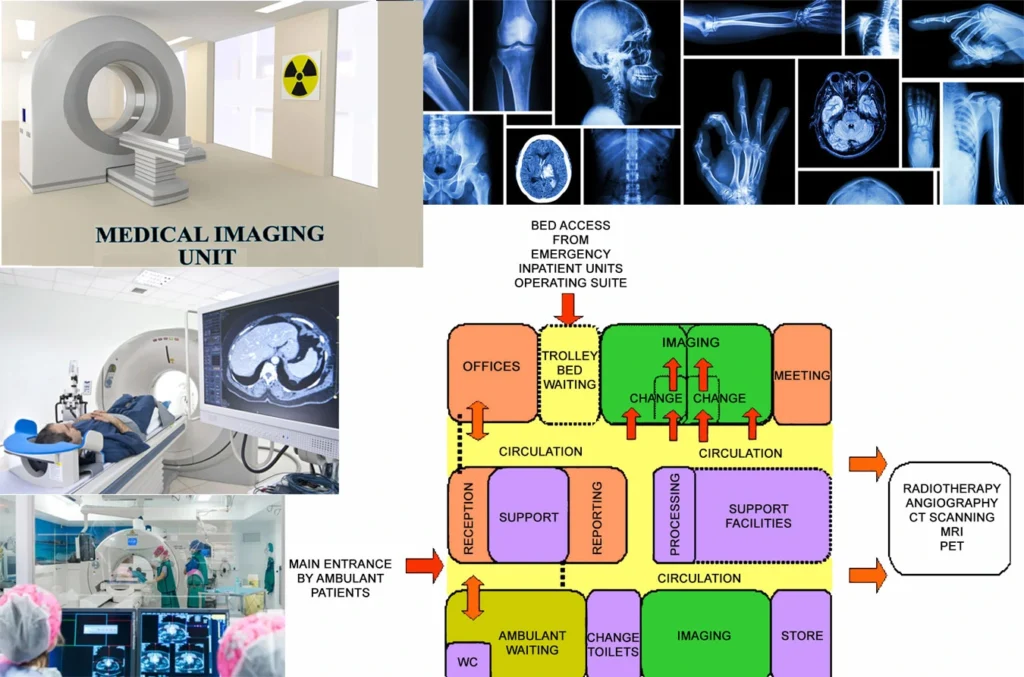
With the advancement of digital imaging and artificial intelligence, medical imaging has become more precise, efficient, and widely accessible. It has significantly reduced the need for exploratory surgeries and has improved patient outcomes through early detection and intervention.
Importance of Well-Designed Medical Imaging Units
A well-designed medical imaging unit is vital for ensuring patient safety, operational efficiency, and compliance with healthcare regulations. Proper design influences not only the quality of diagnostic imaging but also the overall patient experience and staff productivity.
Key benefits of a well-planned medical imaging unit include:
- Optimized workflow – Reduces waiting times and enhances staff efficiency.
- Improved patient comfort – Provides a stress-free environment with minimal noise and radiation exposure.
- Regulatory compliance – Meets safety and accreditation standards such as HIPAA, ACR, and FDA.
- Future readiness – Accommodates emerging technologies such as AI-powered imaging and remote diagnostics.
If you want to know about the Types of slabs or Permeable concrete or Islamic architecture, please click the link.
1) Introduction
The Medical Imaging Unit is a discrete unit of the hospital which provides for General X-ray diagnostic investigations. Depending on the level of service the unit may also provide for diagnostic screening (fluoroscopy), ultrasound, mammography, computed tomography (CT) or interventional radiographic procedures.
The Medical Imaging Unit may be co-located with or incorporate other specialties including Nuclear Medicine, Angiography, MRI, and PET Units.
2) Planning of Design of Medical Imaging Unit
i) Planning Models
The layout of a Medical Imaging Unit should be developed in compliance with manufacturer’s recommendations, because area requirements may vary from machine to machine.
Since technology changes frequently and from manufacturer to manufacturer, rooms should be sized larger to allow upgrading of equipment in the future.
Privatisation of services
Increasingly Medical Imaging services are being delivered as a privately owned and operated service. This option needs to be identified early in the planning process as there may be considerable spatial, design and cost implications.
Off-Site services
In smaller hospitals that cannot justify a full Medical Imaging Unit, access to off-site services is an important consideration in the planning phase, in particular, the selection of the site.
ii) Functional Areas
The Medical Imaging Unit may consist of the following Functional Areas depending on the Operational Policy and service demand:
- Reception and Waiting Areas
- Imaging and screening rooms with access to patient change areas and toilets
- Support areas including preparation areas, storage, disposal and utility rooms
- Film processing areas – both daylight and darkroom areas as required; alternatively, medical imaging may be based on a filmless digital imaging system with its own equipment and storage requirements
- Film storage areas
- Viewing and Reporting areas
- Administrative and Office areas
- Staff Amenities areas including Staff Room, Staff Change Rooms and Toilets and access to Meeting Rooms
Clean utility / preparation areas
The Clean Utility / Preparation Room shall provide for preparation and mixing of contrast media, storage of medications and sterile supplies. The Clean Utility / Preparation Room, if conveniently located, may serve any number of rooms.
The Clean Utility / Preparation Room shall comply with requirements identified in Standard Components – Clean Utility. When preprepared media is used, additional storage shall be provided for the media
Film processing areas
Film processing if required shall be located convenient to the Imaging Rooms and to the quality control area and will normally involve daylight processing equipment. A Darkroom may be provided for specialised processing if required.
The Darkroom, if provided will require special attention to lighting and ventilation. If the Medical Imaging Unit operates with a filmless, digital imaging system, the appropriate areas for image processing and printing will be required according to the type of system installed.
Film storage
For digital imaging applications, there will need to be an area for the PACS (Picture Archiving and Communications System) archive storage units.
A room with cabinets or shelves to file hard copies of patient film shall be provided, located close to the Reception/ administration area. Archived film may be stored outside the Imaging Unit, but must be properly secured to protect films against loss or damage.
General radiology / tomography
Each General will include an upright Bucky stand for chest films. Where volumes are low, OPG, Mammography and Tomography may be added to the General room equipment. This will necessitate a slightly larger room. Tomography is becoming less used with the advent of CT but may be required/ preferred by a Urology service.
The necessary attachments may be incorporated into a General Room. At least one General X-ray room must be sized and located to facilitate transfer of patients from Emergency Unit, if a dedicated room in the EU is not provided.
Orthopantomography (OPG)
OPG is a method of obtaining films of the upper and lower teeth-bearing jaws that supports Trauma, Dental and Facio maxillary services. This equipment may be incorporated into a General Room, a separate bay or within the Dental Unit.
Mammography
Mammography imaging may be included for diagnostic purposes. It should be sized to allow prone positioning for some interventional biopsy procedures. Mammography should be located adjacent to an Ultrasound Room for fine needle biopsies. Change Rooms should be discreet and access to an Interview Room will be required.
Ultrasound
Ultrasound imaging is used in a variety of specialties including Obstetrics, Medicine, Surgery, Cardiology and Vascular Surgery. Ultrasound rooms may be provided within the specialty departments or within the Medical Imaging Unit.
One ultrasound room should be sized to allow for interventional procedures. There must be access to a toilet and drinking water for ultrasound procedures that require the patient to have a full bladder.
Fluoroscopy
Fluoroscopic/radiographic imaging procedures involve administration of contrast media to the patent, serial repositioning of the patient and the timed use of a fluoroscopic imaging system.
The Fluoroscopy room will require a preparation room for barium preparation and an adjacent toilet / shower, accessed from inside the room and from the external corridor.
With the general decline in use of barium contrast studies and advances in equipment technology, fluoroscopy and angiography may be combined in one room. The room must be equipped for anaesthesia.
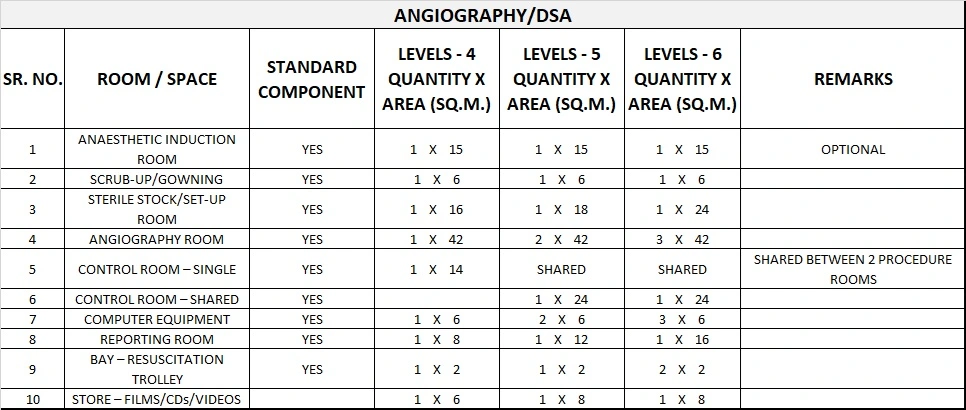
Digital subtraction angiography (DSA)
Simple angiography involves injection of a radiographic contrast agent into blood vessels so that vascular structures are enhanced and revealed together with surrounding bony and soft tissue structures. This procedure is used for simple peripheral studies and can be done on a fluoroscopy table.
With DSA, a contrast agent is administered directly, via a catheter, into an artery close to the area to be examined. The subtraction of a pre-contrast mask suppresses interfering structures from the image so that the arteries become clearly defined.
This process enables a full spectrum of vascular and non-vascular procedures including angiography, angioplasty, arterial and venous stents, biopsy and drainage procedures, and biliary and urologic procedures.
Computerised tomography (CT Scanning)
Refer to the Standard Component for CT Scanning. A Control Room may service 2 rooms. The room may need to be serviced for general anaesthesia. A bed/ trolley bay adjacent to each room is required for staff to observe waiting patients.
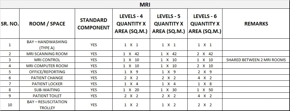
Magnetic resonance imaging (MRI)
MRI will require a dedicated area or suite for access control and protection of/from the magnet (fringe field), and preparation/nursing support areas.
Requirements include:
- Interview room for patient consents and explanations in close proximity
- storage for MRI-compatible (non-ferrous) equipment
- lockers for patient property that may interfere with or be damaged by the magnet such as credit cards and keys.
Careful consideration must be given to the location of the MRI in order to minimise the provision and cost of shielding required including the following:
- MRI should not be located under a helipad or next to a sub-station
- floor / slab must be structurally capable of carrying the weight of the MRI
- good external access is required for the installation of the MRI; a removable side panel may be more cost effective than dismantling a RF shielded door
- room size and shape must be able to contain the 5 Gauss magnetic field with the room and consideration should be given to the needs for future 3T MRIs
- access control needs to be included to ensure only authorised staff enter the MRI room
- locate away from moving ferrous objects which can interfere such as lifts, cars moving through car parks, construction sites
- ensure that emergency equipment such as fire extinguishers and medical gas bottles in the vicinity are not made of magnetic iron.
Endoscopic retrograde choleopancreatography (ERCP)
ERCP is a diagnostic procedure for examination of the biliary and pancreatic ducts system and may be a therapeutic intervention for removal of gall stones etc. It is a procedure used by gastroenterologists, and may be performed in the Medical Imaging Unit or in an Endoscopy Unit.
iii) Functional Relationships
The location of the Medical Imaging Unit, if provided, is variable. Consideration must be given to its proximity to Accident and Emergency, and to the Operating Unit where dedicated in theatre X-ray is not provided.
The requirement for an Outpatient X-ray Service may also dictate where in the facility it is located. In most instances, a compromise between travelling distance for inpatients (minor role) and convenience for outpatients (major role) will be made.
3) Design of Medical Imaging Unit
i) Construction Standards
Special attention is to be given to the following in the design of a Medical Imaging Unit:
- Structural support for equipment including equipment mounted to ceilings
- Level floor for equipment positioning and safe patient movement
- The impact on room space of large diameter electrical cable support tray (in floor and surface mounted)
- Equipment ventilation
- Procedure timing (clocks)
- Task lighting/dimming
- Room blackout, as required.
- Construction Standards for a Medical Imaging Unit include the following:
- Provision for cable trays, ducts or conduits should be made in floors, walls, and ceilings as required.
- Ceiling heights may be higher than normal.
- A lay-in type ceiling should be considered for ease of installation, service, and remodelling.
Standards & codes
Radiological facilities are to comply with relevant State legislation, regulations and statutory requirements.
ii) Environmental Considerations
Acoustics
Acoustic privacy should be provided in all imaging rooms, interview rooms and particularly in reporting areas. Please refer to () “Acoustic Solutions for Healthcare Facilities”
Lighting
Provide indirect and dimmable lighting required in all examination rooms for patient comfort. Ceiling mounted shadowless lighting is required in CT and Angiography imaging rooms.
Privacy
Visual patient privacy is an important consideration to be addressed in the design of imaging rooms and waiting spaces. Privacy screens will be required to imaging and screening rooms.
iii) Infection Control
Hand-washing facilities shall be provided for each Imaging Room, located within or outside the entry to the room. Refer to () – Infection Control: Handwashing Facilities for a discussion on the types of basins suitable for this area.
iv) Space Standards and Components
Rooms shall be sized to suit the design requirements of the equipment to be used, to provide a safe working environment and to allow the effective movement of staff and patients.
Ceiling heights shall suit the equipment, but shall not be less than 3000 mm for ceiling tube mount installations.
Special consideration should also be given to the width and height of doorways to ensure delivery and removal of equipment is not impeded or prevented, and that patient trolley and bed movement is not hampered.
v) Building Service Requirements
Radiation protection
Most Medical Imaging requires radiation protection. Plans and specifications will require assessment for radiation protection by AERB.
The radiation protection assessment will specify the type, location and amount of radiation protection required according to the final equipment selections and layout. Radiation protection requirements shall be incorporated into the final specifications and the building plans.
Communications
Nurse call system
Nurse call buttons shall be located in or near change cubicles, patient-use toilets, showers and at every holding/ recovery bay.
Staff Assist and Emergency Call buttons are required in all Imaging rooms, Holding and Recovery areas Annunciator panels in corridors must be located for optimum viewing.
Voice/ data communicatons
Voice / data installation may include:
- Voice / data cabling for phones and computers
- Dictation system for reporting and / or voice recognition system
- High speed network for digital and CR equipment
- PACS
- Patient or Medical RecordsSystems
- Radiology Information System ideally linked to the Patient Information System
- Conferencing facilities
4) Components of the unit
The Medical Imaging – General Unit will consist of a combination of Standard Components and Non-Standard Components. Provide Standard Components to comply with details in Standard Components described in these Guidelines. Refer also to Standard Components Room Data Sheets and Room Layout Sheets.
i) Non Standard Components
Digital (PACS) Reporting area
Description and Function – PACS reporting areas will include Radiologist workstations for viewing and reporting on procedures using high resolution (LCD) monitors on which images can be manipulated. A minimum of two linked monitors are required, occasionally four screens are provided. In addition to the reporting monitors, a dedicated computer will be required for access to the Patient Information System and a system for dictating reports. .
Location and Relationships – Locate in a quiet are with ready access to the imaging rooms. Several workstations may be located in one room but will need to be visually and acoustically separated.
Considerations – The reporting area will require:
- Ergonomic design of the workstation to accommodate the monitors.
- adequate ventilation and temperature control to individual spaces to minimise risk of monitor failure
- individual cubicle lighting (dimmable)
- Acoustic measures to ensure quality of voice recordings. Please refer to () “Acoustic Solutions for Healthcare Facilities”
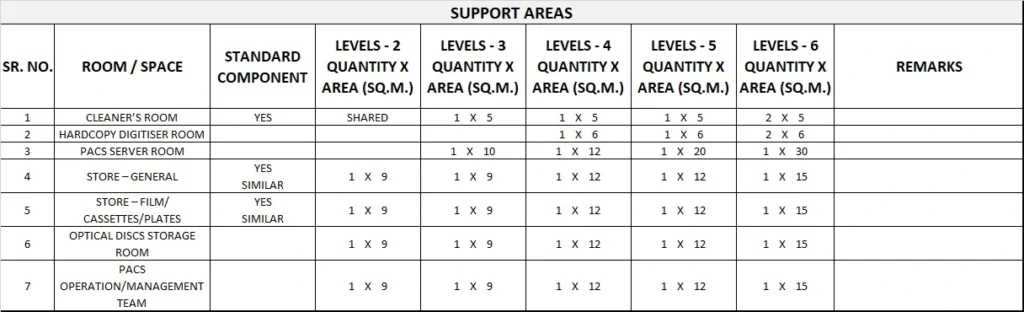
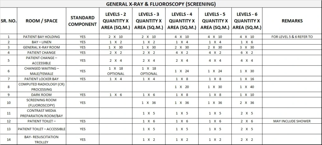
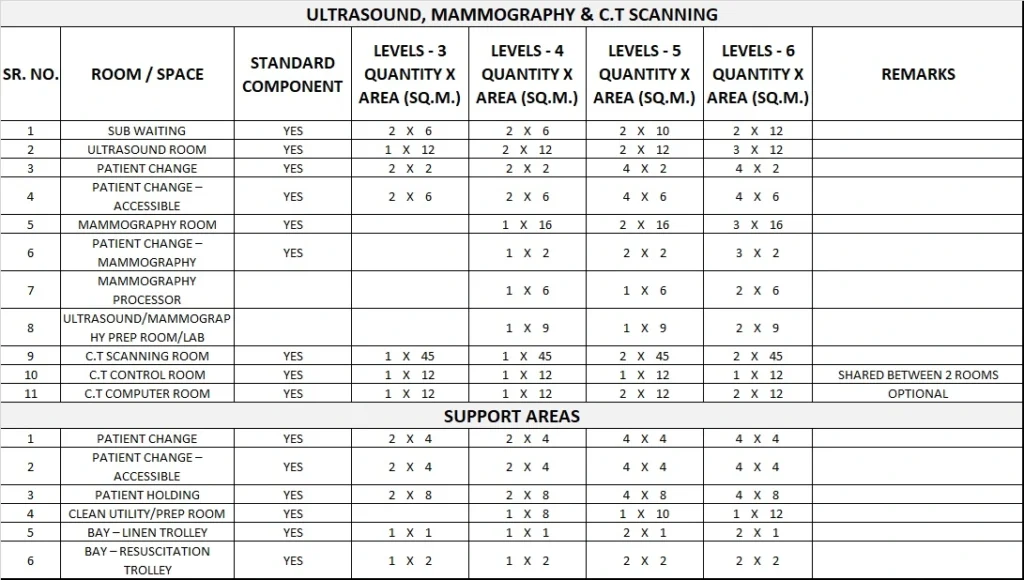
5) Schedule of Accommodation
Medical Imaging-General Generic Schedule of Accommodation
Schedule of Accommodation for a Medical Imaging Unit – General for Level 2-6
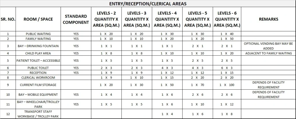
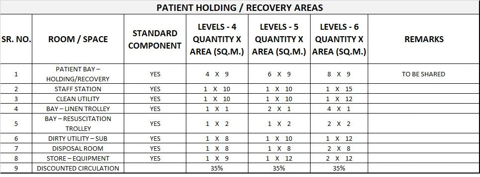
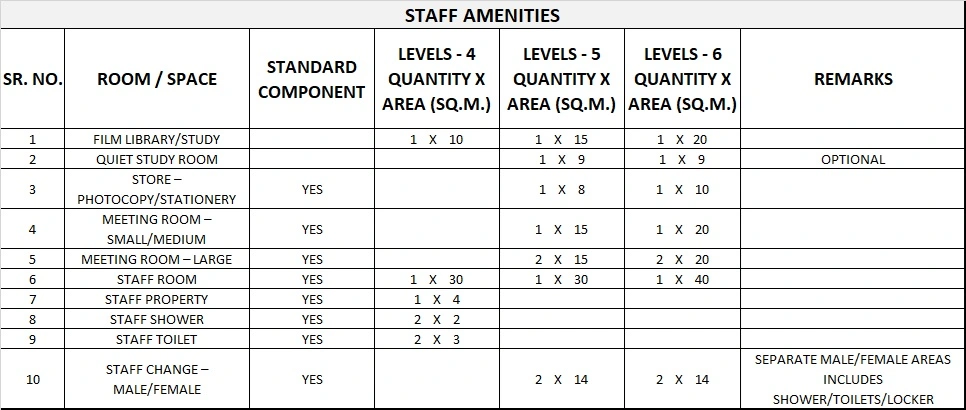
Please note the following:
- Areas noted in Schedules of Accommodation take precedence over all other areas noted in the FPU.
- Rooms indicated in the schedule reflect the typical arrangement according to the Role Delineation.
- Exact requirements for room quantities and sizes will reflect Key Planning Units identified in the service plan and the policies of the Unit.
- Room sizes indicated should be viewed as a minimum requirement; variations are acceptable to reflect the needs of individual Unit.
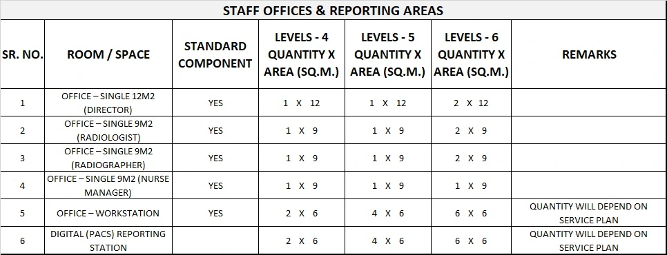
- Office areas are to be provided according to the Unit role delineation and staffing establishment.
- Staff and support rooms may be shared between Functional Planning Units dependant on location and accessibility to each unit and may provide scope to reduce duplication of facilities.
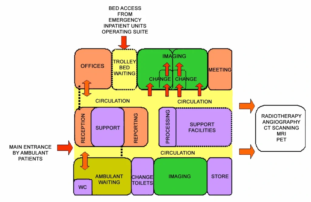
6) Functional Relationship Diagram
Medical Imaging-General Functional Relationship Diagram